


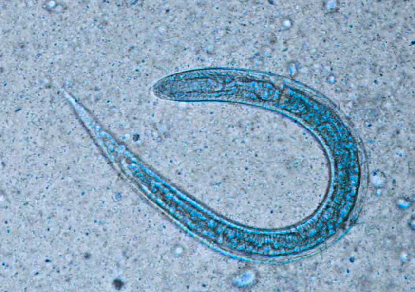
Strongyloidiasis (Strongyloides stercoralis): symptoms, diagnosis and treatment
Strongyloidiasis (Strongyloides stercoralis): symptoms, diagnosis and treatment
Strongyloidiasis is a parasitic infection caused mainly by the nematode Strongyloides stercoralis. An estimated 614 million people worldwide are infected, particularly in tropical and subtropical regions.
Infection occurs when infective larvae present in soil contaminated with human faeces penetrate the skin. The larvae then migrate through the bloodstream to the lungs, ascend the respiratory tract, are swallowed and reach the intestine, where they mature.
One of the most distinctive characteristics of S. stercoralis is its ability to cause autoinfection. Some larvae can re-enter the intestinal wall or the skin of the same host, allowing infection to persist for years or even decades if it is not diagnosed and treated.
Clinical features
Acute and chronic Strongyloides stercoralis infection may be asymptomatic, which can make diagnosis challenging.
When symptoms occur, they mainly affect the skin and gastrointestinal tract and, less frequently, the respiratory system:
- Skin manifestations: an itchy erythematous rash may appear at the site where the larvae entered the skin. A characteristic manifestation is larva currens, a rapidly migrating linear or serpiginous urticarial lesion.
- Gastrointestinal symptoms: abdominal pain, diarrhoea, alternating diarrhoea and constipation, loss of appetite and, occasionally, signs of malabsorption.
- Respiratory symptoms: coughing or wheezing may occur as larvae migrate through the lungs. Severe infections may cause Löffler syndrome, which can be associated with eosinophilia.
Eosinophilia is frequently observed, although it may be suppressed in patients receiving corticosteroids or cytotoxic chemotherapy.
Complications
One of the main reasons why early diagnosis of strongyloidiasis is particularly important is the risk of developing Strongyloides hyperinfection syndrome or disseminated strongyloidiasis.
Severe disease occurs particularly in immunocompromised patients, including people receiving corticosteroids, immunosuppressive therapies or transplantation, as well as patients infected with HTLV-1. A previously asymptomatic infection may progress rapidly after immunosuppressive therapy is initiated.
Potential complications include:
- Hyperinfection syndrome: a marked increase in parasite burden and larval migration through the gastrointestinal and respiratory systems.
- Disseminated strongyloidiasis: larvae spread to organs that are not normally part of the parasite's life cycle, including the central nervous system, liver, heart or skin.
- Bacteraemia and meningitis: disruption of the intestinal mucosa and bacteria carried on migrating larvae can contribute to severe bacterial infections.
- Gastrointestinal complications: ileus, intestinal obstruction, gastrointestinal bleeding, severe malabsorption or peritonitis.
- Respiratory complications: dyspnoea, haemoptysis, pulmonary infiltrates and respiratory failure.
Hyperinfection syndrome and disseminated strongyloidiasis can be life-threatening, particularly in immunocompromised patients.
Diagnosis
The diagnosis of strongyloidiasis can involve parasitological, serological and, in some laboratories, molecular methods.
Direct detection is mainly based on the microscopic identification of larvae in stool samples. However, examination of a single stool sample detects larvae in only around 25% of uncomplicated infections. Repeated examination of concentrated stool samples and specialised techniques such as the Baermann method or agar plate culture can increase diagnostic sensitivity.
Serology for Strongyloides stercoralis is an important tool for supporting diagnosis. Immunoassays can detect anti-Strongyloides antibodies in serum and offer greater sensitivity than microscopic examination of a single stool specimen.
However, antibody testing has some limitations:
- A negative result does not completely exclude infection.
- Cross-reactivity may occur in patients with filariasis or other nematode infections.
- Antibody detection cannot reliably distinguish between current and previous infection.
Antibody levels may decrease following successful treatment, meaning that serology may also contribute to patient follow-up.
Some reference laboratories also use PCR for the detection of S. stercoralis. However, molecular methods have not replaced microscopy and serology as the main diagnostic approaches.
Screening is particularly relevant in people with a history of residence in or travel to endemic areas who also have unexplained eosinophilia, HTLV-1 infection, an upcoming transplant or planned corticosteroid treatment.
Treatment
All patients diagnosed with strongyloidiasis should receive treatment.
Ivermectin is the preferred treatment for uncomplicated infection and has a higher cure rate than albendazole, which may be used as an alternative.
In immunocompromised patients or those with hyperinfection syndrome or disseminated strongyloidiasis, prolonged treatment may be required until parasite clearance has been demonstrated in the samples tested.
Detecting Strongyloides stercoralis infection before starting immunosuppressive therapy is particularly important, as corticosteroid treatment can trigger progression from chronic or asymptomatic infection to severe disease.