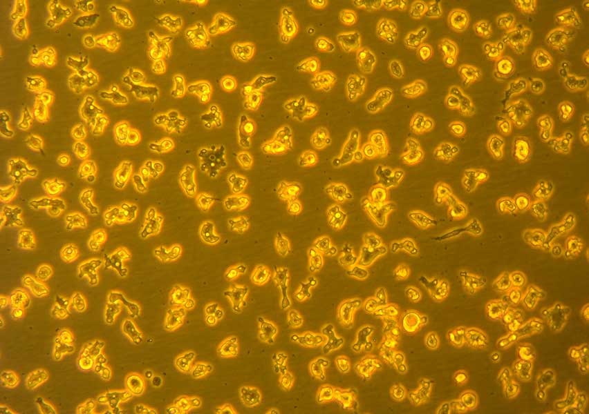
Acanthamoeba
Acanthamoeba are free living amoeba that present two phases in its life cycle: cysts (the resistance form) and trophozoites (the replicating stage). They have been found in varied environments (sea and fresh water, pools, contact lens and dental equipment and solutions, air conditioning systems, animal tissues). It can cause severe keratitis in healthy individuals (particularly contact lens users) and amoebic encephalitis, disseminated disease, or skin lesions in individuals with compromised immune systems.
Acanthamoeba species are classified into three morphologic groups. Group I has large cysts with rounded outer walls (ectocysts) that are clearly separated from the inner walls (endocysts). Group II cysts are smaller, with variable endocyst shapes. Group III cysts are smaller than Group II cysts, with poorly separated walls. The major human pathogens belong to Group II, although A. culbertsoni, from Group III, is also a recognized pathogen. Eight Acanthamoeba species have been isolated as etiologic agents in Acanthamoeba keratitis: A. castellanii, A. polyphaga, A. culbertsoni, A. hatchetti, A. rhysodes, A. lugdunensis, A. quina and A. griffini.
Clinical features: The clinical presentation of Acanthamoeba keratitis, a potentially blinding infection of the cornea, varies greatly. Affected individuals may complain of unilateral foreign body sensation, photophobia, decreased visual acuity, tearing, and pain or redness of the eye. Infection involving both eyes can occur. Pain out of proportion to clinical findings is a classic feature of Acanthamoeba keratitis; however, especially early in the disease, lack of pain does not preclude the diagnosis. Because of similarities to the clinical manifestations of viral, fungal, or bacterial corneal infection, individuals may be misdiagnosed and treated with improper antimicrobial or corticosteroid therapy. Such therapy may initially alleviate symptoms and further obscure the clinical picture and diagnosis.
Diagnosis: The first step in diagnosing Acanthamoeba keratitis is to have a high degree of suspicion, especially in a contact lens wearer with a recent diagnosis of another form of keratitis, such as Herpes simplex virus keratitis, who is not responding to therapy. Diagnosis is made on the basis of clinical picture and isolation of organisms from corneal culture or detection of trophozoites and/or cysts on histopathology. However, a negative culture does not necessarily rule out Acanthamoeba infection. Confocal microscopy and polymerase chain reaction assays to detect Acanthamoeba may also assist with diagnosis.
Treatment: Early diagnosis is essential for effective treatment of Acanthamoeba keratitis. The infection can be difficult to treat due to the resilient nature of the cyst form. Current treatment regimens usually include a topical cationic antiseptic. The duration of therapy may last six months to a year. Pain control can be helped by topical cyclopegic solutions and oral nonsteroidal medications. The use of corticosteroids to control inflammation is controversial. Penetrating keratoplasty may help restore visual acuity.

